Tag Archive for: x-ray

Transforming Medical Imaging with Mini C-Arm X-Ray Technology

What if you could achieve higher diagnostic accuracy while improving patient satisfaction? Enter the world of portable Mini C-Arm X-Ray technology. This innovation offers precision, convenience, and versatility in medical imaging. With trusted providers like ImagPros delivering cutting-edge imaging solutions, equipping your practice has never been easier. 

Evolution of Medical Imaging 

The introduction of the X-Ray in 1895 was the beginning of a long journey for medical imaging. Over the years, advancements such as CTs, MRIs, and larger C-Arm machines have enhanced diagnostic capabilities and supported innovative treatment procedures. However, traditional imaging systems often require significant space, time, and financial investment. 

The mini C-Arm, a more compact and portable evolution of the classic C-Arm, solves these challenges. Designed for precision and accessibility, they are redefining medical imaging’s potential across clinical settings. 

Introducing Mini C-Arm X-Ray Technology 

Mini C-Arm X-Ray systems are portable imaging devices primarily used for real-time imaging of extremities like hands, wrists, ankles, and feet. Their compact size allows medical professionals to bring imaging directly to the patient. If you’re considering buying a C-Arm, the mini C-Arm offers an excellent option with modern features like intuitive designs, easy mobility, and powerful imaging software that produces high-quality visuals. 

Benefits of Mini C-Arm X-Ray Technology 

  • Compact and Portable: Unlike traditional imaging systems, Mini C-Arms fit seamlessly into smaller clinics, operating rooms, and specialized practices, making them a smart choice when buying a C-Arm. 
  • Exceptional Precision: High-quality, real-time imaging offers clarity, improving diagnostic accuracy and procedural efficiency. 
  • User-Friendly: With features like touchscreen monitors and automated image enhancements, Mini C-Arms simplify imaging workflows. 
  • Cost-Effective: These systems require less upfront investment and maintenance than larger imaging equipment, a key factor for those buying a C-Arm.

portable mini c arm
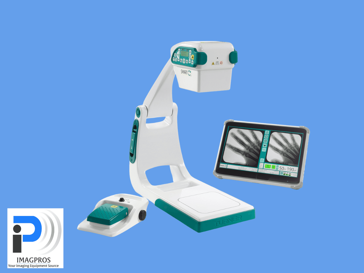
SMART-C® Technology in Clinical Settings 

One standout in Mini C-Arm innovation is SMART-C® technology. Used across medical settings, SMART-C® combines advanced imaging with user-friendly functionality. Its mobility ensures seamless integration into busy practices, allowing medical professionals to focus on patient outcomes. 

SMART-C® Key Advantages 

  • Improved Efficiency: Faster imaging saves time, which allows for higher patient throughput. 
  • Enhanced Accuracy: Crisp, detailed images ensure reliable diagnostic insights. 
  • Patient-Centered Care: Compact designs improve patient comfort by eliminating unnecessary movement in imaging rooms. 

SMART-C® technology represents how Mini C-Arms continues to redefine medical imaging standards. 

Applications of Mini C-Arm X-Ray Technology 

Mini C-Arms have become indispensable in various medical fields. Here’s a closer look at how they’re supporting better diagnostics and treatments in specialized areas. 

Orthopedic Surgery 

Orthopedic procedures often require real-time imaging to assess bones, joints, and implants. Mini C-Arms offer precise visuals, which allow surgeons to make confident, accurate adjustments during procedures. From fracture management to joint replacements, these devices deliver clarity where it matters most. 

Podiatry 

Podiatrists use Mini C-Arms to diagnose foot and ankle conditions. The device provides immediate imaging with no dedicated X-Ray rooms. This reduces patient wait times and accelerates treatment planning, improving overall care quality in podiatric practices. 

Sports Medicine 

Diagnosing sports injuries involves speed and precision, and Mini C-Arms deliver both. They excel at imaging fracture zones, ligament injuries, and common soft tissue conditions in athletes. Additionally, they allow immediate post-operative imaging to confirm procedural success. 

Professional Sports Teams 

Professional sports teams increasingly use mini C-Arms for on-site imaging in training facilities and at live sporting events. Quick diagnoses mean timely treatments, keeping players at peak performance. Their portability makes them invaluable for traveling teams, offering world-class care wherever the game takes them.

Looking Ahead 

Portable Mini C-Arm technology is more than an advancement—it’s a breakthrough in medical imaging. For professionals seeking efficient, versatile, and cost-effective solutions, Mini C-Arms provide superior functionality. If you’re considering buying a C-Arm, ImagPros can provide high-quality imaging solutions, ensuring your clinic has the tools to provide the best possible care.

With ImagPros, you will receive expert guidance, reliable equipment, and seamless implementation. Contact ImagPros today to learn how Mini C-Arms can enhance your practice’s capabilities. 

The Past, Present, and Future of Medical Imaging Devices

How have advances in medical imaging reshaped modern healthcare? Medical imaging has become a critical part of patient care, from detecting hidden diseases to guiding intricate surgeries. But it wasn’t always this way. The evolution of imaging technology has been a remarkable journey—one that began as rudimentary glimpses into the body and has since transformed into a sophisticated diagnostic marvel. Let’s explore the intriguing history, the growing capabilities, and the exciting future of medical imaging devices.

Understanding the History of Medical Imaging Technology 

As we know it today, medical imaging is the culmination of decades of innovation and perseverance. Its importance became evident in the 20th century as scientists and healthcare pioneers pushed the boundaries of what was possible. 

A Century of Rapid Progress in Medical Imaging 

The history of medical imaging is a fascinating journey of innovation and discovery, marked by milestones that revolutionized how we understand, diagnose, and treat diseases. These advances have transformed medicine, providing clinicians unprecedented ways to visualize the human body.

  • 1895: The Discovery of X-Rays 

Wilhelm Röntgen’s discovery of X-rays was a pivotal moment in medical science. By uncovering the ability to see inside the human body without invasive surgery, Röntgen opened the door to modern diagnostics. Initially met with awe and curiosity, X-rays quickly became a staple, effectively detecting fractures, infections, and foreign objects. Despite its immense potential, long exposure times, and a lack of safety protocols, which prompted further research into making it safer and more efficient, limited early X-ray technology.

  • 1970s-1980s: Emergence of CT Scans and MRIs 

The advent of Computed Tomography (CT) and Magnetic Resonance Imaging (MRI) redefined medical imaging. In the early 1970s, CT scans allowed clinicians to capture cross-sectional images of the body using a combination of X-rays and computer processing. This provided unparalleled detail for evaluating complex brain, chest, and abdomen structures. 

MRIs, widely available in the 1980s, took imaging further using magnetic fields and radio waves, enabling soft tissue visualization without radiation exposure. MRIs provide detailed insights into conditions such as tumors, neurological disorders, and joint abnormalities, making them invaluable in oncology, neurology, and orthopedics. These technologies improved diagnostic accuracy and drove the development of specialized fields like interventional radiology.

  • 1990s-Present: Integration of Digital Imaging 

The shift from analog to digital imaging in the 1990s marked another transformative era. Digital Radiography (DR) and Picture Archiving and Communication Systems (PACS) revolutionized image capture, storage, and sharing. Digital imaging eliminated the need for physical film, significantly reducing storage costs and environmental impact while enhancing image clarity and resolution. 

Integrating advanced software and artificial intelligence (AI) expanded digital imaging’s potential. AI algorithms can now assist radiologists by identifying anomalies, suggesting diagnoses, and streamlining workflows. The global adoption of telemedicine has allowed digital imaging to reach patients in remote or underserved areas, bridging gaps in healthcare access.

Building the Foundation for the Future 

Each of these breakthroughs has advanced healthcare and laid the groundwork for the innovations we see today. From portable ultrasound devices to AI-driven imaging systems, the evolution of medical imaging continues to make diagnostics faster, more accurate, and more accessible. As we look to the future, technologies like 3D imaging, molecular imaging, and augmented reality-guided procedures hold immense promise, ensuring that the story of medical imaging is far from complete.

Medical Imaging Devices

The Role of Deep Tissue Imaging 

Deep tissue imaging sits at the core of modern medicine. Enabling detailed visualization of tissues, organs, and systems provides unmatched clarity that traditional methods cannot achieve. 

Why It’s Important 

Deep tissue imaging isn’t just about seeing the unseen. It’s about understanding the human body’s complexities to deliver accurate diagnoses and effective treatments. This technology guides physicians through procedures like tumor resections, organ transplants, and trauma surgeries with greater precision than before. 

For instance:

  • Cancer Detection: PET scans pinpoint abnormal tissue growth early, improving survival chances. 
  • Neurological Disorders: MRI scans map intricate brain activity, aiding the treatment of conditions like epilepsy or multiple sclerosis.
  • Joint Health: Ultrasound-guided imaging delivers precise diagnostics for musculoskeletal injuries, helping athletes and patients recover faster. 

Deep tissue imaging doesn’t just uncover problems—it offers a window into solutions, improving patient outcomes and quality of life. 

The Present: Advances in Medical Imaging Technology 

Today’s medical imaging devices are more advanced than ever, packed with cutting-edge capabilities that only a few decades ago felt like science fiction. 

The State of Modern Medical Imaging 

Innovative imaging technologies now drive decision-making across hospitals and clinics worldwide. Modern CT scanners deliver high-resolution images in seconds, while portable ultrasounds bring imaging to remote or intensive care environments. Systems built with AI capabilities enable healthcare professionals to optimize workflows, reduce diagnostic times, and enhance accuracy. 

At the center of this evolution are companies like ImagPros, providing turnkey imaging solutions to meet the diverse demands of healthcare providers. Their dedication to customer satisfaction ensures access to high-quality imaging devices that integrate seamlessly with modern practices, including refurbished and custom-built systems tailored to specific needs. 

Expert Service and Customer Satisfaction 

With over 50 years of combined experience, ImagPros knows that optimal imaging solutions require more than just devices—they need support. From equipment implementation and training to state compliance and financing, our end-to-end services reflect a commitment to excellence in the field. 

Looking Toward the Future 

The future of medical imaging will be even more exciting as emerging trends and technologies advance diagnostics and treatments to previously unimaginable heights. 

Next-Generation Technologies 

  • AI Integration 

Artificial intelligence is at the forefront of innovation, rapidly analyzing imaging data to enhance diagnostic accuracy. AI can identify conditions like heart disease or lung cancer earlier and more precisely than traditional methods allow. 

  • 3D and 4D Imaging 

Three-dimensional imaging is already reshaping surgical planning, while 4D imaging focuses on real-time dynamic motion, aiding fields like cardiology and orthopedics. 

The Potential Impact on Healthcare 

These advancements promise faster diagnostics, personalized treatments, and reduced healthcare costs. For example, AI-powered imaging could lessen the burden on radiologists, allowing medical professionals to focus on critical cases and deliver more timely care. For patients, this means better health outcomes and fewer diagnostic errors. 

Medical Imaging as the Backbone of Modern Healthcare 

Medical imaging has become an indispensable part of healthcare, bridging the gap between symptom onset and life-saving treatment. From its humble beginnings to its current state as a sophisticated technology field, imaging devices have redefined the art of medicine. 

Looking ahead, companies like ImagPros will continue to play a vital role, leading the charge with innovative solutions that empower healthcare providers. With the right tools and partnerships, the future of medical imaging holds incredible promise. 

Interested in learning how high-quality imaging devices can support your practice?

Contact ImagPros to explore state-of-the-art imaging solutions and see how we can transform your diagnostics and patient care approach.

Refurbished C-Arm Cost: How Old Is Too Old When Buying Used?

What if you could cut costs without sacrificing reliability? Well, you can. The answer is the refurbished C-Arms. The balance between ensuring performance and managing the C-Arm cost is challenging, but trusted providers like ImagPros ensure that refurbished systems deliver quality and compliance you can trust whether you purchase a 5 or 15-year old C-Arm. 

The Vital Role of C-Arms in Modern Healthcare

The C-Arm has become an essential tool in medical imaging, offering unmatched precision and flexibility. Its ability to deliver live, real-time imaging makes it indispensable for a wide range of procedures, including orthopedic surgeries, pain management, cardiovascular imaging, and more. With continuous technological advancements, C-Arms enhance surgical outcomes by increasing accuracy and reducing procedure times. This improves patient safety and boosts operational efficiency for healthcare providers. Whether investing in a new or refurbished system, a reliable C-Arm can significantly elevate the quality of care.

Key Features of C-Arms

New C-Arms

New C-Arms have cutting-edge flat panel detectors that deliver exceptional image clarity, making them ideal for high-precision orthopedics, cardiology, and surgery procedures. They often integrate seamlessly with AI-powered imaging systems, providing real-time analysis and automated adjustments to improve diagnostic accuracy. These systems also prioritize user convenience, featuring intuitive touchscreen controls, advanced mobility for flexible use in clinical settings, and reduced radiation exposure for improved safety.

Refurbished C-Arms

Refurbished C-Arms offer a cost-effective solution for healthcare facilities without compromising on performance. Thoroughly restored and upgraded to meet current standards, these systems include reliable image intensifiers that ensure consistent image quality. Enhanced with updated software and hardware, refurbished models are well-suited for a variety of procedures, such as pain management, urology, and vascular surgery. They provide high functionality comparable to new systems but at a fraction of the cost.

Choosing the Right C-Arm

Understanding the distinct features and benefits of new versus refurbished C-Arms is crucial for making informed decisions. Both options allow you to optimize your healthcare investments while maintaining a high standard of patient care. 

For more information on how C-Arms can transform your medical practice, contact ImagPros today.

Understanding the Refurbishing Process 

Refurbished C-Arms aren’t just cleaned and resold. The process is a meticulous series of inspections and upgrades designed to restore systems to near-new condition. Here’s an overview of what happens during refurbishment, as practiced by reputable providers like ImagPros: 

  • Inspection & Decontamination: Each system undergoes a full review for wear, functionality, and potential damage. Following this, all parts are rigorously decontaminated with antimicrobial cleaners, ensuring the equipment meets strict hygiene standards. 
  • Part Replacement & Repairs: Components that fail to meet OEM specifications are repaired or replaced. This includes batteries, power supplies, cables, monitors, and structural elements like locking fixtures and brakes. 
  • Calibration for Precision: Settings such as collimators, image intensifiers, and beam alignment are fully calibrated to ensure imaging accuracy and performance reliability. 
  • Thorough Testing & Quality Control: The system is tested under real-world conditions and checked against state-specific compliance requirements. Only after passing these tests is it approved for resale. 

By the end of this process, the refurbished system is as close to new as possible while being significantly more affordable. 

refurbished c-arm
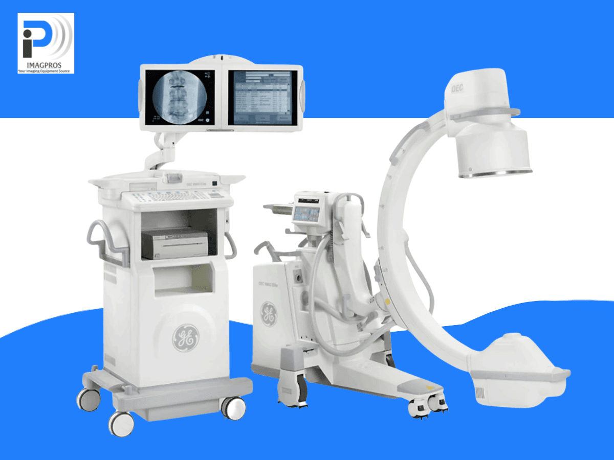
Why Refurbished C-Arms Often Focus on OEC 

OEC C-Arms are one of the most commonly refurbished systems due to their reputation for durability, reliability, and performance. Known for their high-quality image intensifiers and precise fluoroscopic capabilities, OEC systems remain a favorite among medical facilities worldwide. Their robust build and modular design make them ideal candidates for refurbishment, as they can be easily repaired, upgraded, and recalibrated to meet modern standards. 

Additionally, OEC systems have widespread availability of replacement parts, making the refurbishment process more efficient while ensuring long-term serviceability. When refurbished, OEC C-Arms maintain their impressive imaging quality and diagnostic accuracy, offering a like-new experience at a fraction of the cost. These factors make OEC systems a go-to choice for refurbishers and buyers alike. 

Meeting Industry Standards and Regulatory Requirements 

A common concern when buying used or refurbished equipment is whether it meets industry standards. All refurbished C-Arms from trusted providers like ImagPros comply with FDA, ISO, and CE regulations, ensuring they are safe, reliable, and legally approved. 

Compliance with these standards protects patient safety and your practice. By adhering to these requirements, you meet legal obligations and build patient trust in your services. 

Warranty, Service Support, and Reliability 

One key factor when purchasing medical equipment is the post-purchase support offered. Reputable refurbishers like ImagPros provide a comprehensive warranty covering parts and services for each unit. Buyers also gain access to dedicated service teams ready to assist with maintenance and repairs, ensuring minimal downtime for your operations. 

Reliable warranties and service packages ensure that buying refurbished doesn’t compromise peace of mind. These safeguards often rival and sometimes even exceed those offered with new units. 

Technology Upgrades and Performance 

Refurbished C-Arms are not outdated relics—they often feature upgraded technology that keeps them competitive with newer models. Older systems can be retrofitted with modern software and hardware enhancements, such as improved imaging algorithms or flat screen monitors.

These upgrades ensure your refurbished system matches current standards and typically delivers the same level of diagnostic accuracy as a brand-new unit without the hefty price tag. 

For comparison, OEC C-Arms, known for their reliability and versatility, are commonly refurbished. Features like their high-quality image intensifiers and precise fluoroscopic capabilities are preserved and often enhanced for long-term use through quality refurbishing. 

The Cost Advantage of Used and Refurbished C-Arms 

When balancing finances and equipment quality, refurbished systems are hard to beat. Refurbished options offer significant cost savings compared to buying new units—upfront and operational costs. 

While the exact C-Arm cost will vary depending on the brand, model, and refurbished, facilities can often save anywhere from 30% to 50% of the price of a new unit. These savings can be crucial for practices operating with tight budgets or prioritizing financial flexibility. 

Savings don’t stop at the initial purchase. With warranties and modern upgrades, operational expenses remain predictable, while performance remains dependable.

Maximize Results with ImagPros 

Investing in a refurbished C-Arm is not just a budget-friendly choice—it’s a smart business decision when reliability is ensured. So, the question isn’t “how old is too old? Instead, it’s “Who is handling the refurbishment process? 

ImagPros specializes in helping healthcare facilities access high-quality refurbished medical imaging equipment that complies with industry standards and delivers trusted performance. Ask us about our wide selection of C-Arms that range in age and capability, today! 

Our expertise, from detailed refurbishing processes to reliable warranties and cost efficiencies, ensures you find the right solution for your practice. 

Contact ImagPros today to explore our refurbished C-Arms and learn more about how these systems can provide quality and value for your healthcare facility. 

Portable X-Ray Technology Revolutionizing Healthcare

Picture a healthcare landscape where speed, accuracy, and convenience merge for seamless patient care. This is not a distant future but the present reality with portable X-Ray technology. At the forefront, ImagPros offers innovative solutions that are reshaping diagnostics in healthcare. We explore the evolution of digital imaging in healthcare and why it’s paving the way forward.

Portable X-Ray Technology

Portable X-Ray systems offer a flexible and efficient solution for diagnostic imaging, particularly in environments where mobility is crucial. These systems are designed to deliver high-quality imaging while being portable and easy to operate, making them indispensable in a variety of medical settings.

Key components of a portable X-Ray system include:

  • Portable X-Ray Generator – Produces the X-rays needed for diagnostic imaging.
  • DR Panel (Direct Radiography Panel) – Captures X-ray images digitally with precision.
  • PC (Personal Computer) – Processes, stores, and manages the radiographic images.
  • Specialized Software – Enhances image quality and helps streamline workflows.

To offer even greater versatility, optional accessories can be incorporated into these systems:

  • X-Ray Table – Assists with positioning patients during the imaging process.
  • Wall DR Holder – Allows for upright imaging, useful for chest X-rays and other vertical examinations.
  • Handheld Devices – Provide maximum portability for imaging in tight or unconventional spaces.

The combination of these essential and optional components enables portable X-Ray systems to meet a wide range of diagnostic needs. Their adaptability, reliability, and ease of use ensure that healthcare professionals can perform imaging efficiently and effectively, no matter where the patient is located.

The Evolution from Analog to Digital Imaging 

The shift from traditional analog to digital X-Rays represents a significant advancement in medical imaging, streamlining processes and enhancing image quality. Key benefits include: 

  • Reduced wait times 
  • Instant access to high-resolution images 
  • Faster diagnosis and treatment 
  • Easy storage and retrieval of patient records 
  • Improved data management 
  • Cost efficiency through reduced physical storage and consumables 

This transition allows healthcare facilities to utilize resources more effectively and increase productivity.

Digital X-Ray and the Future of Healthcare

Advancements in digital X-Ray technology are transforming medical imaging. These systems use artificial intelligence and machine learning to improve diagnostic accuracy and speed. AI algorithms analyze X-Ray images to identify patterns and anomalies that may be missed by the human eye, aiding radiologists in making informed decisions and minimizing errors. Machine learning also enables predictive analytics, providing insights into potential health issues early on. Additionally, digital X-Ray systems are increasingly integrated with electronic health records (EHRs), facilitating seamless data sharing and enhancing patient management.

portable x-ray technology

Digital Imaging’s Impact on Rural and Remote Areas

Access to quality healthcare can be challenging in rural and remote areas. Portable X-Ray machines help by providing diagnostic capabilities to underserved regions. This accessibility allows patients to receive timely imaging services, reducing the need to travel long distances.

Healthcare providers benefit from improved diagnostics without high infrastructure costs. Mobile units can serve multiple locations, maximizing resource efficiency and expanding care reach. Digital imaging also supports telemedicine, enabling specialists to review X-Rays remotely. This collaboration enhances diagnostic accuracy and aids in making informed treatment decisions, regardless of location.

Addressing Common Concerns About Portable X-Rays 

While the benefits of portable X-Ray technology are evident, some healthcare professionals may have concerns regarding image quality, radiation safety, and system integration. Advancements in sensor and Detector technology ensure that portable units provide high-quality images comparable to stationary systems. Meanwhile, advanced filtering techniques minimize radiation exposure for patient safety. Integration with existing healthcare infrastructure is seamless, as digital systems support various formats and communication protocols, allowing portable X-Rays to improve current workflows.

ImagPros: Leading the Way in Portable X-Ray Solutions

ImagPros is dedicated to delivering innovative portable X-Ray systems that enhance diagnostic accuracy and streamline healthcare operations. By choosing ImagPros, you gain access to cutting-edge technology and unparalleled support, empowering your practice to provide superior patient care.

Join the imaging care revolution and transform your facility with ImagPros today!

Demystifying C-Arm Fluoroscopy: 4 Essential Facts You Should Know

In the complex world of medical imaging, making an informed decision about equipment like versatile C-Arm machines can often feel overwhelming. Whether you’re a seasoned healthcare professional or new, we’re here to help. At Imagpros LLC, we’ve compiled four crucial aspects you need to know about C-Arm fluoroscopy.

Understanding C-Arm Machines and Their Functionality

C-Arm machines have revolutionized the field of medical imaging with their exceptional versatility and mobility. These groundbreaking devices are extensively used in various medical procedures, including surgical, orthopedic, and complex cardiac interventions. The name “C-Arm” comes from the distinctive C-shaped arm connecting the X-ray source and detector

This design enables unparalleled flexibility and maneuverability during imaging procedures. This remarkable feature allows the C-Arm machine to navigate around the patient, capturing precise and detailed images from various angles. C-Arm devices have become indispensable tools in modern medicine. 

These advanced technologies empower healthcare professionals with superior imaging capabilities, enabling precise diagnoses and guiding effective treatments.


Contact us today to upgrade your imaging capabilities with a new C-Arm!

The Mechanism and Key Components of Mobile C-Arm Machines


A mobile C-Arm is a crucial medical device that operates through three key components: an X-ray generator, an image intensifier, and a monitor. The X-ray generator, powered by advanced technology, emits high-energy X-rays that penetrate the patient’s body, providing detailed images for diagnostic and surgical purposes. These X-rays then pass through the patient and are captured by the image intensifier, which amplifies and enhances the image quality. 

The intensified image is transmitted to the monitor, where healthcare professionals can observe the live feed in real time. This real-time visualization empowers medical practitioners to make accurate and timely decisions during complex procedures, ensuring optimal patient care and safety.

C-Arm Fluoroscopy
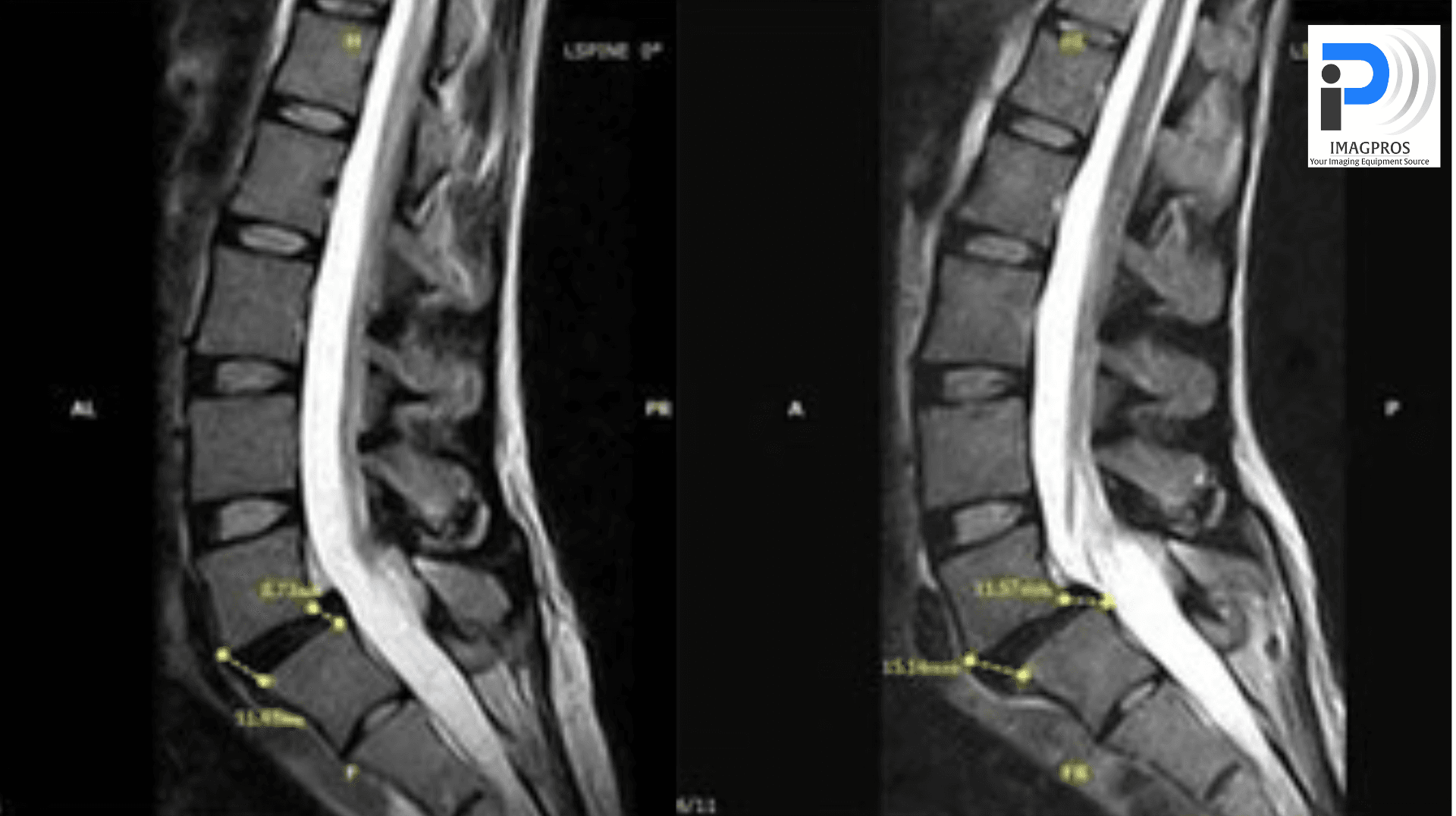
Comparing C-Arm with Radiography or Fixed Fluoroscopy Machines

The differentiating factor between C-Arm machines and radiography or fixed fluoroscopy machines lies in their exceptional mobility and flexibility. These attributes enhance their functionality and provide greater convenience and versatility in various medical settings. Unlike their fixed counterparts, C-Arms can be moved and adjusted to capture images from different angles without moving the patient. This makes them ideal for dynamic procedures that require real-time imaging.

Top 3 FAQs When Buying a C-Arm Machine

Before investing in a C-Arm machine, it’s essential to consider a few critical factors:

  1. Ideal X-Ray Generator Power: The power of the X-ray generator is crucial as it affects the image quality. Higher power generators provide clearer images, especially for larger patients or dense areas. It’s worth noting that some Brand New systems have features that can produce clear images even with lower-powered generators.
  2. Machine Dimensions & Maneuverability: Consider the dimensions and maneuverability of the C-Arm depending on your facility’s space. Some models are more compact and easier to maneuver, making them suitable for smaller areas.
  3. Tube Type – Stationary Anode or Rotating Anode: The type of tube can significantly impact the life and performance of the C-Arm. Especially when looking at older generation C-Arms, machines with rotating anodes typically have a longer lifespan and provide better cooling than those with stationary anodes.

At Imagpros LLC,, we are dedicated to providing high-quality turn-key imaging solutions. Our team of experts is always ready to guide you through selecting the most suitable C-Arm machine for your needs. 

For anything imaging-related, consider the pros—consider Imagpros.

Safeguarding Against Radiation: Effective C-Arm Machine Strategies

In the ever-evolving medical imaging field, innovation continues to amaze. Mobile C-Arm Systems have revolutionized examining the human body and conducting procedures. However, new technological advancements bring challenges, like ensuring radiation safety for healthcare professionals. Let’s dive into this intriguing topic.

The Core of Modern Imaging: What is a C-Arm X-Ray Machine?

A C-Arm is an X-ray machine with a distinctive C-shaped arm connecting the X-ray source and detector. This design allows for flexible positioning, producing detailed images from various angles. The devices emit X-rays from one end of the C-Arm, which pass through the body, and the detector captures them on the opposite side. This imaging technique gives clinicians detailed views of internal structures, helping accurate diagnoses and precise treatments.

The New Horizon in Mobile C-Arm Machines

Contemporary Mobile C-Arm Systems are significant advancements in medical technology. They offer clear images, user-friendly interfaces, and adaptability in both stationary and mobile setups. The latest models prioritize enhanced radiation safety, reducing operator risk.

Breakthroughs in technology have introduced 3D imaging capabilities, providing physicians with a comprehensive view of the patient’s anatomy. This is invaluable in complex surgical procedures that require precision.

Find the right C-Arm for your practice with ImagPros!

radiation
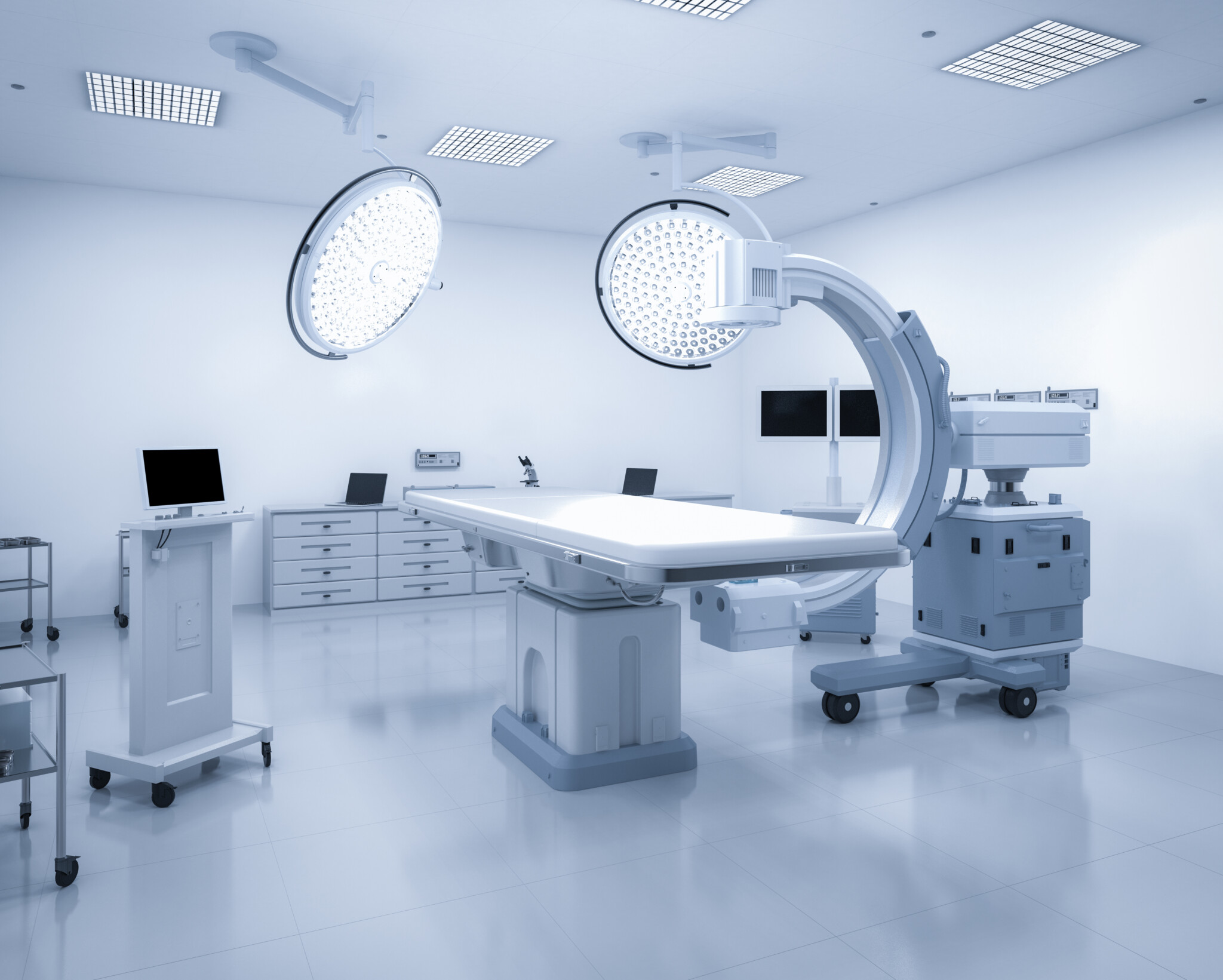
The Challenge of Radiation Safety in Mobile C-Arm Systems

Healthcare professionals have valid concerns about radiation exposure from a Mobile C-Arm. The radiation dose from a fluoroscopy procedure can vary based on factors like duration, distance, and shielding. 

Modern Mobile C-Arm machines have safety features to minimize radiation exposure.

These include:

Prioritizing Safety: Shielding Doctors from Radiation Exposure

In medical imaging, healthcare professionals’ safety is crucial. Innovative systems are essential but involve radiation exposure. Therefore, professionals must have comprehensive safety knowledge.

  • Comprehensive Training: Thorough device training is vital for a safer work environment. Professionals learn to minimize exposure and operate systems effectively. This training demonstrates our commitment to delivering top-tier technology and safety expertise.
  • Protective Clothing: Protective clothing is crucial for workplace safety. Lead aprons and thyroid shields provide protection during procedures, reducing risk. Healthcare professionals can perform their duties confidently and safely with these essential items.
  • Adherence to ALARA Principles: Upholding ALARA (As Low As Reasonably Achievable) principles is crucial for radiation safety. It shows our commitment to minimizing radiation doses and reducing the release of radioactive substances into the environment.
  • Regular Maintenance Checks: Regular maintenance checks are crucial for our safety protocol. They optimize system function, preventing radiation leaks or excessive emissions. Our attention to detail sets us apart, ensuring our systems are safe and effective.
  • Advanced Models with Integrated Safety Features: We recommend selecting advanced models with built-in safety features. Modern devices have mechanisms to minimize radiation exposure, ensuring that the benefits of these innovative systems outweigh the risks.

Embracing the Future

Mobile C-Arm Systems innovations reshape the medical landscape, bringing us closer to a future of accessible and secure diagnosis and treatment. As we explore and refine these technologies, it’s our responsibility to ensure their effective and safe usage.

At ImagPros, we provide top imaging solutions focusing on your safety. 

Contact ImagPros to learn more now!

C-Arm Machines: Key Factors in Choosing a C-Arm for Surgery

As medical professionals, ensuring the success of surgical procedures is of utmost importance. One crucial factor in achieving this success is having the right equipment to best maximize your practice. To make an informed decision when selecting a C-Arm for surgery, it’s essential to consider several key factors. This blog provides valuable insights into these factors, ultimately helping you choose the right C-Arm machines to benefit your medical practice and your patients.

Understanding C-Arms

A C-Arm is an imaging device that employs X-ray technology to generate real-time, high-resolution images during surgical procedures. The name originates from the C-shaped arm that connects the X-ray source and detector. This unique design allows for effortless movement and precise positioning around the patient.

To operate C-Arm machines, the X-ray source emits beams that pass through the patient’s body. On the opposite side, the detector or Image Intensifier captures these beams and converts them into digital images. These images are then displayed on a monitor, providing surgeons real-time visual guidance throughout the procedure.

Medical professionals commonly use C-Arms in various fields, such as orthopedic, vascular, and cardiac surgeries while also playing a significant role in pain management and emergency procedures. C-Arms improve patient outcomes and enhance surgical precision by offering accurate and real-time imaging.

Image Quality and Resolution

The primary purpose of a C-Arm machine is to provide clear and accurate images during surgery. High-resolution images allow surgeons to see intricate details and make precise movements, reducing the risk of complications and improving patient outcomes. Therefore, image quality and resolution should be at the top of your list when evaluating different systems.

ImagPros can help you choose a brand-new C-Arm or an affordable refurbished model!

Size and Mobility

C-Arms come in various sizes, from compact and mobile units to more extensive, fixed systems such as Cath Labs. The size and mobility of the device depend on the type of surgeries performed and the space in the operating room. Smaller, mobile C-Arms are ideal for facilities with limited space and offer the flexibility to move between different rooms or departments. These models typically have a smaller footprint and may even feature a monitor on top of the C-Arm instead of having a separate monitor cart, further conserving space.

Ease of Use and Ergonomics

Surgeries can be lengthy and demanding, so choosing a user-friendly and ergonomic C-Arm machine is essential. An easy-to-use system will reduce the learning curve for staff and minimize fatigue during long procedures. Look for a system that is counterbalanced with intuitive controls, easy-to-read displays, effortless movements from AP to lateral, and adjustable components to accommodate various users and patient positions.

Compatibility with Other Equipment

When selecting a C-Arm, it’s crucial to consider its compatibility with other equipment in your facility, such as monitors, surgical tables, and imaging software. A seamless integration with your existing setup will prevent workflow disruptions and help you avoid additional expenses for upgrades or replacements.

Modern C-Arm machines typically feature digital output capabilities, allowing for easy connection to PACS (Picture Archiving and Communication System) and EMR (Electronic Medical Record) software. However, some older models may only offer PACS connectivity and not support exporting images in the JPG format required by certain EMR software.

To ensure smooth integration and optimize the efficiency of your medical practice, verify the compatibility of the chosen C-Arm unit with your specific EMR software. If necessary, explore additional hardware or software solutions that can bridge any gaps between the C-Arm machine and your existing infrastructure.

Service and Support

Finally, consider the level of service and support the manufacturer or distributor provides. A reliable C-Arm is an investment in your practice, so it’s crucial to have access to prompt technical help, maintenance, and repairs when needed. Inquire about training programs and resources available to help your staff become proficient in using the new equipment.

Making the Right Choice

Choosing the right C-Arm for surgery involves evaluating image quality, size, mobility, ease of use, compatibility, and service support. By weighing these aspects, you can select a system that meets your needs and enhances your surgical procedures’ efficiency and effectiveness.

Ready to explore your options and find the perfect C-Arm machine for your practice?


Contact our team at ImagPros today to discuss your requirements and discover the best solutions for your surgical needs.

A Comprehensive Guide to C-Arm X-Ray Fluoroscopy

As a leading imaging equipment provider, we at ImagPros understand the importance of having the right tools for the job. Let’s unlock the essentials together! This comprehensive guide dives deep into C-Arm X-ray fluoroscopy, covering everything from its definition to its various components and applications.

What is a C-Arm Fluoroscopy

A C-Arm machine is a specialized medical imaging device that uses X-rays to produce real-time images of the body’s internal structures. Its name comes from the arm’s distinctive “C” shape, which allows the X-ray source and image intensifier to be positioned on opposite sides of the patient. This design provides unparalleled flexibility and maneuverability, enabling healthcare professionals to get high-quality images from virtually any angle.

What is an X-Ray Image Intensifier?

In any C-Arm fluoroscopy, the X-ray image intensifier is the central component that converts X-ray energy into visible light. The image intensifier produces top-quality images with minimal distortion and noise. A viewer can see real-time photos on a screen as the camera captures light.  Some newer models now feature a flat detector instead of an image intensifier, as well.

c-arm x-ray fluoroscopy


Who Uses a C-Arm?

Many healthcare professionals use C-Arm machines, including orthopedic surgeons, interventional radiologists, and pain management specialists among many others. They are particularly well-suited for use during surgical procedures, as they allow doctors to visualize the area being treated with no invasive techniques. Some typical applications include:

  • Orthopedic surgery, such as joint replacements or spinal fusions etc…
  • Interventional radiology, including angioplasty and stent placement etc…
  • Pain management for targeted injections, nerve blocks, kyphoplasty, RF Ablation, stimulator implants, etc…

What Are the Components of a C-Arm?

Several key components make up a C-Arm system, including:

  1. X-ray generator: This device generates X-rays to create images of the body’s internal structures.
  2. C-Arm: The characteristic “C” shaped arm houses the X-ray source and image intensifier , allowing them to be positioned opposite each other.
  3. Image intensifier: As discussed earlier, this component converts X-ray energy into visible light, which is then captured by a camera.
  4. Monitor: The monitor displays real-time images, enabling healthcare professionals to view and assess the patient’s condition during a procedure.

These are just a few of the essential components that make up a C-Arm system. With their incredible versatility and powerful imaging capabilities, these machines can be invaluable tools in the hands of an experienced healthcare provider.


If you want to upgrade your imaging equipment, contact us today, and let’s get started!

Reasons to Use a C-Arm System

There are several reasons a C-Arm fluoroscopy system is an invaluable tool for healthcare professionals:

  • Real-time imaging: C-Arm machines provide real-time images, allowing doctors to make adjustments and confirm the success of a procedure as it’s happening.
  • Minimally invasive: By providing clear, detailed images, C-Arm machines enable doctors to perform procedures with minimal invasiveness, reducing recovery time and improving patient outcomes.
  • Versatility: A C-Arm machine’s unique design allows for unparalleled positioning flexibility, making it suitable for various applications.
  • Efficiency: With high-quality images at the touch of a button, C-Arm machines streamline the imaging process, saving time and resources.

Ready to unlock the power of a C-Arm X-ray machine for your practice?

Contact ImagPros at in**@******os.com to learn more about our range of state-of-the-art imaging equipment and expert support services for C-Arm fluoroscopy. Together, we’ll ensure you have the tools to provide the best care for your patients.

Shielding Plan Consideration

A shielding plan is a document that details the materials and designs used to isolate radiation in an area. It outlines which materials should be used, where they should be placed, and how effectively they prevent radiation from entering or leaving the room. Shielding plans can also specify additional safety protocols for radiation-related activities, such as using appropriate clothing and protective equipment. Each state has its own set of regulations and guidelines for shielding plans, so it’s essential to understand the specific details of your location.

The ImagPros Promise

ImagPros is proud to offer a customized consulting program tailored to Turn-key solutions when purchasing our equipment. Our team of experts will help you create an effective shielding plan compliant with state regulations. We’ve done the research, so you don’t have to—our five-step process allows us to provide you with a detailed list of rules that need to be followed, along with suggestions on which materials will provide the best protection. Every X-ray and fluoroscopy sale comes with a free shielding plan that will be state compliant—we won’t leave you alone to figure it out on your own. We strive to provide the best protection for our customers and their businesses.

Our Customized Plans

Each business is unique, and we design your shielding plan based on the size and layout of your facility. We consider traffic patterns, room dimensions, and materials used in construction to create a plan that meets your needs. If any changes need to be made to an existing structure or activity spaces, we’ll provide detailed recommendations for implementing these. We also consider the radiation in your area and create shielding plans that protect staff and customers from unnecessary exposure.

What Do We Need for Requirements?

We need specific information about your facility to create a shielding plan, including the room’s layout and windows. We’ll also need to know the floor you’re on and the material used between floors (typically steel beam with 2 inches of concrete). Finally, we’ll need to know how many procedures you expect to be performed each week and the amount of fluoroscopy/x-ray time per patient.

1. Drawing of the room (with Length x Width)5. Anything above/below?
2. What is on each side of the wall?6. What is the material between floors (typical material is steel beam with 2 inches of concrete)
3. Any Windows?7. How many procedures expected per week?
4. What floor are you on?8. How much fluoro time per patient?

Shielding Plan

We also consider lead thickness requirements. For Fluoroscopic Procedure Rooms, 1/32 inch lead is the general requirement. For typical Diagnostic Stationary X-ray Rooms, 1/16 inch lead may be needed. We offer a Lead Door with reinforced hinges to support the door’s weight and can supply alternative options in our Action Plan. Our lead can be purchased in 4 feet by 7 feet sheets for installation in wall construction.

We look forward to helping you protect your employees and customers in the most effective way possible.


Learn More