Tag Archive for: imaging

Portable X-Ray Technology Revolutionizing Healthcare

Picture a healthcare landscape where speed, accuracy, and convenience merge for seamless patient care. This is not a distant future but the present reality with portable X-Ray technology. At the forefront, ImagPros offers innovative solutions that are reshaping diagnostics in healthcare. We explore the evolution of digital imaging in healthcare and why it’s paving the way forward.

Portable X-Ray Technology

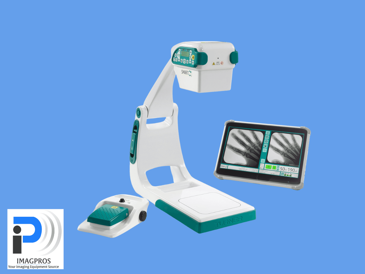
Portable X-Ray systems offer a flexible and efficient solution for diagnostic imaging, particularly in environments where mobility is crucial. These systems are designed to deliver high-quality imaging while being portable and easy to operate, making them indispensable in a variety of medical settings.

Key components of a portable X-Ray system include:

  • Portable X-Ray Generator – Produces the X-rays needed for diagnostic imaging.
  • DR Panel (Direct Radiography Panel) – Captures X-ray images digitally with precision.
  • PC (Personal Computer) – Processes, stores, and manages the radiographic images.
  • Specialized Software – Enhances image quality and helps streamline workflows.

To offer even greater versatility, optional accessories can be incorporated into these systems:

  • X-Ray Table – Assists with positioning patients during the imaging process.
  • Wall DR Holder – Allows for upright imaging, useful for chest X-rays and other vertical examinations.
  • Handheld Devices – Provide maximum portability for imaging in tight or unconventional spaces.

The combination of these essential and optional components enables portable X-Ray systems to meet a wide range of diagnostic needs. Their adaptability, reliability, and ease of use ensure that healthcare professionals can perform imaging efficiently and effectively, no matter where the patient is located.

The Evolution from Analog to Digital Imaging 

The shift from traditional analog to digital X-Rays represents a significant advancement in medical imaging, streamlining processes and enhancing image quality. Key benefits include: 

  • Reduced wait times 
  • Instant access to high-resolution images 
  • Faster diagnosis and treatment 
  • Easy storage and retrieval of patient records 
  • Improved data management 
  • Cost efficiency through reduced physical storage and consumables 

This transition allows healthcare facilities to utilize resources more effectively and increase productivity.

Digital X-Ray and the Future of Healthcare

Advancements in digital X-Ray technology are transforming medical imaging. These systems use artificial intelligence and machine learning to improve diagnostic accuracy and speed. AI algorithms analyze X-Ray images to identify patterns and anomalies that may be missed by the human eye, aiding radiologists in making informed decisions and minimizing errors. Machine learning also enables predictive analytics, providing insights into potential health issues early on. Additionally, digital X-Ray systems are increasingly integrated with electronic health records (EHRs), facilitating seamless data sharing and enhancing patient management.

portable x-ray technology

Digital Imaging’s Impact on Rural and Remote Areas

Access to quality healthcare can be challenging in rural and remote areas. Portable X-Ray machines help by providing diagnostic capabilities to underserved regions. This accessibility allows patients to receive timely imaging services, reducing the need to travel long distances.

Healthcare providers benefit from improved diagnostics without high infrastructure costs. Mobile units can serve multiple locations, maximizing resource efficiency and expanding care reach. Digital imaging also supports telemedicine, enabling specialists to review X-Rays remotely. This collaboration enhances diagnostic accuracy and aids in making informed treatment decisions, regardless of location.

Addressing Common Concerns About Portable X-Rays 

While the benefits of portable X-Ray technology are evident, some healthcare professionals may have concerns regarding image quality, radiation safety, and system integration. Advancements in sensor and Detector technology ensure that portable units provide high-quality images comparable to stationary systems. Meanwhile, advanced filtering techniques minimize radiation exposure for patient safety. Integration with existing healthcare infrastructure is seamless, as digital systems support various formats and communication protocols, allowing portable X-Rays to improve current workflows.

ImagPros: Leading the Way in Portable X-Ray Solutions

ImagPros is dedicated to delivering innovative portable X-Ray systems that enhance diagnostic accuracy and streamline healthcare operations. By choosing ImagPros, you gain access to cutting-edge technology and unparalleled support, empowering your practice to provide superior patient care.

Join the imaging care revolution and transform your facility with ImagPros today!

Transforming Medical Diagnostics with Flat Panel Detector C-Arms

Medical imaging is undergoing a revolution. With Flat Panel Detector C-Arms, the future holds promise for unprecedented clarity and efficiency in diagnostics. These technological advancements will redefine how healthcare professionals approach diagnostics, promising a future where precision and speed are paramount.

Understanding the Shift: Flat Detector C-Arm Technology 

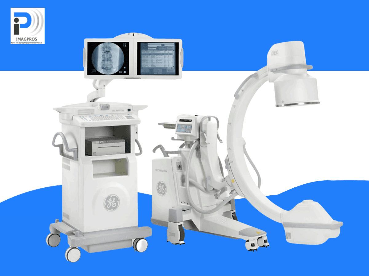
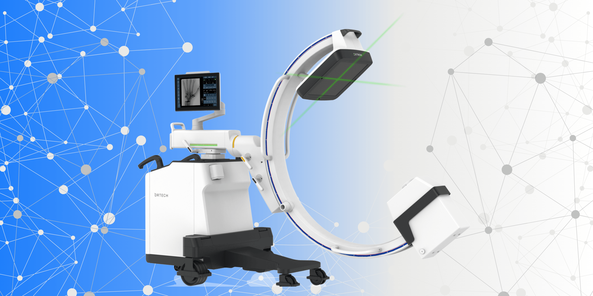
Flat Detector C-Arms (FPD C-Arms) are transforming the medical imaging landscape. Unlike traditional C-Arms, which use image intensifiers, FPD C-Arms employ flat-panel detectors. This ongoing shift results in improved image quality, reduced radiation exposure, and increased flexibility in capturing detailed anatomical structures. 

The Evolution from Traditional C-Arms to Flat Detector C-Arms 

While traditional C-Arms have served the medical community well, their limitations are becoming increasingly apparent. FPD C-Arms offer superior spatial resolution and dynamic range. As the transition from analog to digital imaging continues, the medical community increasingly captures every detail more accurately, facilitating better diagnostic decisions and positioning FPD C-Arms to become the standard of care. 

Primary Applications and Benefits Over Traditional Systems 

FPD C-Arms are versatile and are being adopted across various medical specializations, including orthopedics, cardiology, and vascular interventions. They provide significant benefits, such as lower radiation doses, enhanced image clarity, and faster processing times, making them essential tools in routine and complex procedures and setting the stage to become the preferred choice in medical imaging.

Flat Detector C-ArmFuture Innovations with FPD C-Arms

The field of medical imaging is continually evolving, and FPD C-Arms are at the forefront of this evolution. Innovations in AI, machine learning, and real-time imaging will further enhance these devices’ capabilities.

Current Advancements and Their Implications

Current advancements include AI-driven image analysis, which aids in quicker and more accurate diagnosis. Improvements in detector sensitivity and image processing algorithms mean that FPD C-Arms can now capture images in previously challenging conditions, such as in patients with high body mass index (BMI).

Predictions for the Future of Imaging with FPD C-Arms

Future innovations will likely focus on integrating FPD C-Arms with other diagnostic tools, creating a seamless imaging ecosystem. Real-time data sharing between devices could enable instantaneous feedback during surgical procedures, enhancing outcomes and reducing procedure times.

The Role of Imaging Professionals in Driving These Innovations

Imaging professionals play a crucial role in adopting and optimizing these technologies. By leveraging their expertise, they implement and continually refine advancements to meet the dynamic needs of medical diagnostics.

Impact on Medical Diagnostics

FPD C-Arms will shift the landscape of medical diagnostics by enhancing accuracy, speed, and patient outcomes. Their impact is already being felt in various medical fields.

Enhancing Accuracy and Speed in Diagnostics

The high-resolution images provided by FPD C-Arms allow for more precise diagnostics. This reduces the likelihood of misdiagnosis and ensures that treatment plans are based on the most accurate data. Faster imaging also means that patients spend less time under radiation.

Real-World Cases of Improved Patient Outcomes and Cost-Effectiveness

In real-world scenarios, FPD C-Arms have significantly enhanced patient outcomes. For example, detailed imaging helps surgeons make precise incisions in orthopedic surgeries, reducing recovery times and improving surgical success rates. The efficiency gained from quicker diagnostics translates to cost savings for medical facilities.

Potential for Integrating FPD C-Arms in Various Medical Specializations

The versatility of FPD C-Arms makes them suitable for a wide range of medical specializations. From pain management, orthopedic to neurology, these devices are indispensable in providing high-quality care.

The Competitive Edge with ImagPros

ImagPros commits to pushing the boundaries of medical imaging. ImagPros sells FPD C-Arms with the latest technology, ensuring healthcare providers can access the best tools available.

Commitment to Innovation and Quality

Innovation and quality are steadfast commitments for ImagPros. Its new and refurbished FPD C-Arms incorporate innovative technology, ensuring medical professionals can perform their duties precisely and confidently.

Features and Support That Differentiate ImagPros’ FPD C-Arms

ImagPros’ FPD C-Arms features advanced noise reduction, low-dose radiation technology, and user-friendly interfaces. Their customer support is unparalleled, providing training and ongoing help to ensure users can fully leverage their equipment’s capabilities.

Explore the Future of Imaging Technology

In summary, the future of medical diagnostics is bright, with FPD C-Arms leading the way. These devices offer unparalleled accuracy, speed, and efficiency, making them an essential tool for any modern medical facility.

Discover how ImagPros can help you elevate your imaging capabilities. Contact us today for a personalized consultation and explore our range of FPD C-Arms designed to meet your needs.

Enhancing Patient Care with Diagnostic Ultrasound Machines

In the fast-paced world of medical diagnostics, staying ahead with reliable and advanced tools is crucial. The diagnostic ultrasound machine offers unparalleled imaging capabilities that enhance patient care by providing accurate, real-time insights into various medical conditions. In the following guide, we’ll explore how the device operates, its diverse applications, and the numerous benefits it can bring to your practice.

Understanding the Basics of Ultrasound Technology

Ultrasound technology employs high-frequency sound waves to capture detailed images of the body’s internal structures. Unlike X-rays or CT scans, which use ionizing radiation that can be harmful with excessive exposure, ultrasounds use sound waves, making them a safer alternative for patients and healthcare providers. This method is helpful for imaging soft tissues, including organs, muscles, and blood vessels, without the risks associated with radiation.

The machine’s processing unit and display system collaborate to interpret and visualize the data effectively, ensuring accurate diagnostic results. The critical component includes the ultrasound transducer, which emits sound waves and captures the returning echoes from internal structures, converting them into diagnostic visual images. This seamless integration of the processing unit, display system, and the primary component—the transducer—ensures precise medical imaging.

The Role of the Ultrasound Transducer

Now, let’s explore the ultrasound transducer in greater detail. This handheld device transmits and receives sound waves, transforming electrical energy into sound waves that penetrate the body. These waves bounce off various tissues and structures, returning echoes that the transducer captures. The ultrasound machine then converts these echoes into visual images, offering real-time insights into the patient’s condition displayed on a monitor.

Capturing Images: The Process Explained

When performing an ultrasound scan, the technician places the transducer on the patient’s skin and typically applies a conductive gel to ensure optimal sound wave transmission. As the transducer moves over the skin, it sends sound waves and receives the returning echoes. The machine processes these echoes to create detailed images of internal organs, tissues, and blood flow.

For certain procedures, doctors can insert the transducer into the body, such as in transvaginal or transrectal ultrasounds, to get a closer view of specific internal structures. This capability enhances the diagnostic potential of ultrasound technology, making it versatile for various medical applications.

Ready to review the different options for your practice? Contact ImagPros at 248-951-9020!

Different Uses of Diagnostic Ultrasound Machines

Diagnostic ultrasound machines’ versatility extends beyond essential imaging, making them indispensable tools across multiple medical fields. From obstetrics to cardiology, they offer critical insights that aid in accurate diagnosis and effective treatment plans.

Obstetrics and Gynecology

One of the most well-known diagnostic ultrasound machines used in obstetrics and gynecology. Ultrasound scans are instrumental in monitoring fetal development during pregnancy, allowing physicians to track growth, detect anomalies, and assess the mother and baby’s health.

Cardiology

In cardiology, ultrasounds evaluate the heart’s structure and function. Echocardiograms, a type of ultrasound, provide detailed images of the heart’s chambers, valves, and blood vessels, aiding in diagnosing and managing cardiac conditions.

Musculoskeletal Imaging

Musculoskeletal ultrasound is invaluable for diagnosing injuries and conditions affecting muscles, tendons, ligaments, and joints. It offers a non-invasive way to visualize soft tissue structures, making it an essential tool for sports medicine and orthopedic practices.

Abdominal Imaging

Ultrasound scans commonly examine abdominal organs, including the liver, kidneys, pancreas, and gallbladder. They help detect abnormalities such as tumors, cysts, and stones, providing critical information for treatment planning.

Vascular Imaging

Doppler ultrasound technology enables the visualization of blood flow in arteries and veins. It is crucial for diagnosing vascular conditions like deep vein thrombosis, arterial blockages, and aneurysms, improving patient outcomes through timely intervention.

diagnostic ultrasound machines

External vs. Internal Ultrasound Procedures

Understanding the distinction between external and internal ultrasound procedures can help healthcare professionals choose the method for each diagnostic requirement. External ultrasounds are typically non-invasive and involve placing the transducer on the skin’s surface to capture images of underlying structures. Abdominal, obstetric, and musculoskeletal imaging often require their use.

In contrast, internal ultrasounds involve inserting the transducer into the body, offering a more direct view of specific internal structures. Procedures like transvaginal and transrectal ultrasounds fall under this category and are highly useful for specific diagnostic needs. For example, transvaginal ultrasounds provide detailed images of the uterus and ovaries, essential for gynecological assessments.

Now that we understand the types of ultrasound procedures let’s learn more about the critical considerations for choosing the right diagnostic ultrasound machine for your practice.

External Ultrasound

Medical professionals conduct external ultrasounds by placing the transducer on the skin’s surface. They widely use this method to image superficial structures such as the thyroid, breast, and musculoskeletal system. It is non-invasive and comfortable for patients, making it suitable for routine examinations.

Internal Ultrasound

Internal ultrasounds involve inserting a specialized transducer into a body cavity. Examples include transvaginal ultrasounds for assessing reproductive organs and transrectal ultrasounds for evaluating prostate health. These procedures provide higher-resolution images of internal structures, offering more accurate diagnostic capabilities for specific medical conditions.

Advanced Features of Diagnostic Ultrasound Machines

Now that we have discussed the basic and specific applications, let’s discuss the advanced features that enhance the capabilities and efficiency of diagnostic ultrasound machines.

Tissue Harmonic Imaging (THI)

Tissue Harmonic Imaging improves image quality by reducing noise and enhancing signal clarity. This technology provides clearer, more detailed images essential for accurate diagnosis.

3D and 4D Imaging

3D and 4D ultrasound technologies offer dynamic views of anatomical structures. 3D imaging constructs three-dimensional images, while 4D imaging adds the element of time, providing real-time visualization of moving structures, such as a beating heart or a developing fetus.

Doppler Imaging

Doppler imaging includes color, power, pulsed-wave, and continuous-wave Doppler. These techniques visualize blood flow and vascular structures, aiding in the assessment of circulatory health and the detection of blood flow abnormalities.

Elastography

Elastography assesses tissue stiffness, helping diagnose conditions such as fibrosis and tumors. It provides additional information on tissue properties, complementing traditional ultrasound images and improving diagnostic accuracy.

Discover How an Ultrasound Machine Can Boost Your Practice

Diagnostic ultrasound machines have revolutionized medical imaging by providing safe, non-invasive, and versatile solutions for various clinical needs. Their ability to capture high-quality images and support diverse applications makes them indispensable in modern healthcare settings.

Contact ImagPros today to learn more about how diagnostic ultrasound machines can enhance your practice and improve patient care. 

Together, let’s shape the future of healthcare.

A Guide to Mobile C-Arm Machines in Modern Medicine

In the dynamic and often high-stakes world of healthcare, staying at the forefront of technological advancement is crucial. These innovative tools enhance the quality of care and broaden the spectrum of what is possible in surgical and interventional procedures. Among these tools stands the mobile C-Arm, a compact yet powerful imaging device that has redefined the standards of intraoperative visualization and patient care. Below, we’ll explore the expert uses and benefits that mobile C-Arm machines bring to the table, revolutionizing medical practices across the globe.

Amplifying or Intensifying the Light Signal

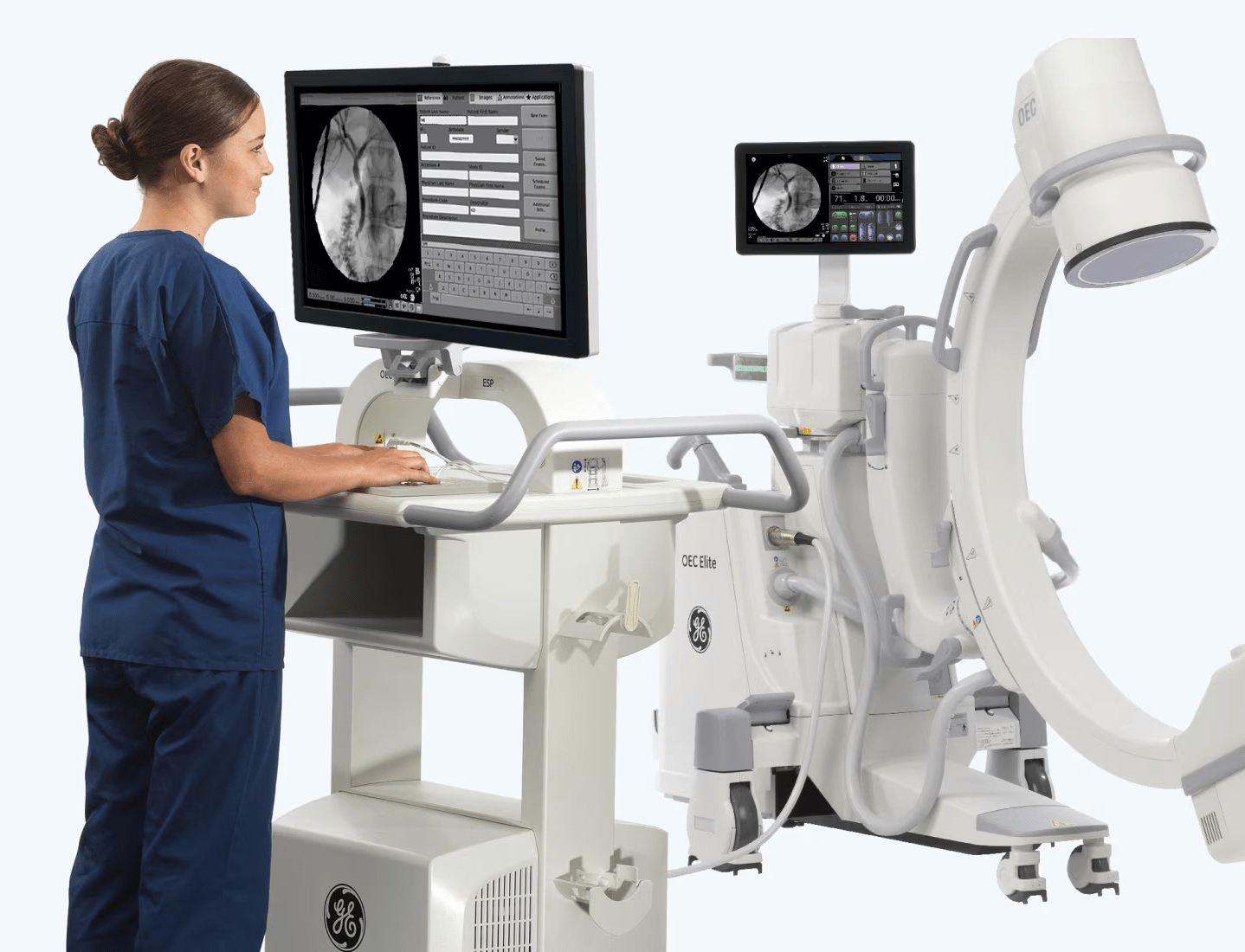
When precision and clarity are non-negotiable, mobile C-Arms deliver where others fall short. These devices boast an image intensifier—an intricate piece of technology—that does what its name suggests: significantly amplifies the light signal created by X-Ray beams. This process transforms obscure, internally cast shadows into detailed, real-time images that guide surgeons through intricate maneuvers. Imagine the line between a blurry, garbled radio transmission and a crystal-clear conversation—the difference an intensifier in a mobile C-Arm can provide during critical operations.

Clinical Need for Mobile C-Arm Imaging

The Mobile C-Arm is not a one-size-fits-all solution—it’s a precise tool for applications as diverse as the healthcare landscape. From orthopedic surgery, which aids in the accurate placement of implants, to emergency room traumas, which necessitate immediate and effective diagnosis, mobile C-Arms is a clinical mainstay. In these situations, speed, flexibility, and reliability are essential—attributes that the mobile C-Arm possesses in abundance. Its ability to pivot, arc, and tilt, coupled with swift image capture and data transfer, allows for a fluid, unimpeded flow in the surgical protocol.

Fluoroscopy in a C-Arm

Within the context of a mobile C-Arm, fluoroscopy becomes the keystone of dynamic, real-time imaging. A type of medical imaging that shows a continuous X-Ray image, the fluoroscopy capabilities of the C-Arm are instrumental in procedures that demand movement and monitoring, like those in Angiography or pain management. Not only does this technology aid in visualizing the structure and movement of internal structure organs with no invasive surgery, but it also plays an integral role in ensuring safe and precise medical intervention.

mobile c-arm machines

Functioning of the Image Intensifier

The image intensifier is the heart of a mobile C-arm’s imaging process and operates on sophisticated principles. The input phosphor screen converts X-Rays into visible light signals upon impact. Because of the conversion process within the image intensifier tube, which involves acceleration and multiplication, the high-intensity light becomes focused and captured by the output phosphor screen. This intensification process allows for detailed images, while reducing the risk of overexposure to X-Rays and minimizing the radiation dose to the patient and medical staff.

Working with an Image Intensifier

The intricate workings of the image intensifier are a marvel of medical engineering. The path from X-rays to visible light follows a concerted series of events that occur within fractions of a second. More than just a simple amplification, the working C-Arm image intensifier ensures that surgical teams receive immediate and continuous feedback, aligning with the fluid nature of their procedures. Through this process, mobile C-Arms offer a marriage of precision and speed unmatched by conventional imaging methods.

Image Location in the C-Arm Intensifier

The strategic placement of the image intensifier within the C-Arm enhances its usability and effectiveness. Its location ensures surgeons have an unobstructed view of the operating field, fostering efficiency and reducing the necessity for cumbersome repositioning. This saves valuable time and streamlines the surgical process, allowing healthcare professionals to focus on what truly matters—the patient.

Closing Thoughts

The mobile C-Arm is a testament to human ingenuity’s capability in healthcare technology. It is a beacon of advance that continues to drive medical progress, offering enhanced imaging capabilities that positively impact patient outcomes. With benefits ranging from reduced procedural times to lower radiation doses, mobile C-Arm machines are not just tools but partners in the mission to provide exceptional care. 

Now is the moment to unlock the full potential of mobile C-arm machines. Contact ImagPros and discover how this revolutionary imaging solution can completely change your healthcare practice.

The Evolution of Ultrasound Technology in Medicine

In the stirring symphony of medical progress, few instruments have played as versatile a role as ultrasound technology. Pioneered decades ago, ultrasound’s metamorphosis from a mere echo to the detailed images that shape modern medical diagnostics is a testament to human ingenuity. This post will dissect the instrumental progression of ultrasound devices, from their sonar roots to the crisp, comprehensive imaging they offer today.

The Echo of Ultrasound’s Origins

They had a more austere beginning before ultrasound devices adorned the clinical halls. Researchers repurposed SONAR technology to examine the human body in the early 20th century. Early adopters in the medical community used simple A-mode technology to detect anomalies. Still, it was in the 1950s that B-mode, or brightness-mode, ultrasound arrived, adding a visual dimension to the sound waves. Ultrasound’s foray into the medical field was nascent but promising, akin to a young virtuoso discovering their talent’s reach.

Ultrasound TechnologyUltrasounds for Medical Use: A Panacea for Imaging Needs

Ultrasound’s utility in medicine is as expansive as it is essential. From detecting gallstones to imaging the beating heart, ultrasound has become a frontline diagnostic tool. Unlike X-rays or MRIs, ultrasound uses non-ionizing forms of radiation, making it exceptionally safe for prenatal screenings. Recent technological advances have fine-tuned ultrasound capabilities, allowing for real-time, high-resolution imaging of soft tissues and organs, cementing its place as the physician’s stethoscope of the 21st century. Although ultrasound is a potent diagnostic tool, it has its constraints. The operator’s proficiency significantly affects the imaging quality, leading to diverse results. 

The Visual Symphony of 3D and 4D Ultrasounds

While traditional 2D ultrasounds are the workhorses of diagnostics, 3D and 4D imaging add an element of depth and time that was previously unattainable. Akin to viewing a stunning landscape in various lights and seasons, 3D ultrasounds offer a three-dimensional glimpse of the fetus, organs, and tissues, enhancing the detail and the sense of space. Integrating the fourth dimension, time, into 4D ultrasounds offers a dynamic view of moving structures, transforming fetal evaluations and bonding moments for expectant parents.

These imaging technologies impact obstetrics and gynecology, where the visual element is deeply personal. Cardiologists also reap the benefits, as 3D and 4D ultrasounds provide essential insights into the structure and function of the heart, guiding them through the symphony of cardiac care with unparalleled precision.

ImagPro’s Crescendo in Ultrasound Evolution

Your medical practice’s orchestra deserves the best players, and at ImagPros, we understand that the right tools can elevate the performance and the outcome. Our company stands at the forefront of ultrasound technology, offering state-of-the-art equipment that embodies the decades-long evolution we’ve detailed here. We ensure your diagnostic skills are exceptional by putting customers first and staying updated on industry advancements.

For consultation on choosing the proper ultrasound devices for your practice, our team at ImagPros is your dedicated conductor, orchestrating a seamless transition to the most current imaging solutions.

Contact us today to play a part in the ongoing evolution of ultrasound technology in medical care!

The Difference Between Refurbished & Used Medical Imaging Equipment

High-quality imaging technology is the lifeblood of any modern healthcare facility, providing crucial diagnostic capabilities that can mean the difference between life and death. As medical professionals and administrators weigh their options, a critical decision arises: should they opt for refurbished or used imaging equipment? This post aims to demystify the distinction between the two and provide clarity for those navigating this critical choice.

The Nuances of “Used” Imaging Equipment

The term “used” often conjures preconceived notions of wear and tear, signaling caution to potential buyers. However, “used” doesn’t denote poor quality in medical imaging equipment. Used medical imaging equipment has been previously owned and operated in a healthcare facility.

Pros and Cons For Medical Facilities

At a glance, the immediate pro is cost savings. Medical facilities, especially those operating under tight budgets, can leverage used equipment to gain the technology at a fraction of the cost of new machinery. This, in turn, can mean better diagnostic capabilities for a broader patient base.

There are risks associated with used equipment. Its history might involve unknown technical failures or maintenance issues. This lack of transparency can lead to unforeseen costs and the potential for equipment downtime, which, in healthcare contexts, can have serious consequences.

Cost-Saving Benefits and Risks

While the allure of a lower sticker price is strong, it’s paramount for medical professionals to discern that the long-term costs, including those related to maintenance and lost operational hours, could outweigh the upfront savings. Used medical imaging equipment is best approached with comprehensive due diligence, which could involve demands for service records, warranty details, and on-site inspections.

medical imaging equipmentThe Refurbishing Process Unveiled

In contrast, refurbishing involves rigorous processes to restore used imaging equipment to a state where it can perform at or near original manufacturer equipment (OEM) specifications.

Critical Steps Involved in Refurbishing

Visualize a meticulous sequence of steps to breathe new life into each piece of equipment. From initial inspection that dissects the machine’s current condition to replacement and recalibration of parts, the process is a testament to the dedication required to transform from used to rejuvenated status.

Focus on Reliability and Quality Assurance

The essence of medical imaging equipment refurbishing lies in its commitment to reliability and quality assurance. Manufacturers like Imagpros offer transparency that used equipment cannot, documenting every aspect of the refurbishing process to ensure their products’ operational safety and effectiveness. Warranties and after-sales support that provide peace of mind to healthcare providers often underscore this level of detail.

The Benefits of Refurbished Imaging Equipment

Choosing refurbished medical imaging equipment can be a strategic move for both the short and long term.

Improved Performance and Longevity

Refurbished machinery typically outperforms standard “used” equipment, sometimes even matching the capabilities of new models thanks to updated parts and stringent testing. As a result, refurbished machinery provides diagnostic accuracy and gives you peace of mind, knowing that manufacturers take pride in building equipment to last.

Enhanced Safety Features and Compliance

In the fast-evolving world of healthcare technology, staying compliant with regulations is non-negotiable. Refurbished equipment often incorporates the latest safety features and technological advancements, ensuring that a healthcare facility is always at the forefront of patient safety and industry standards.

Cost-Effectiveness Compared to New Equipment

The financial benefit remains a powerful draw, with refurbished equipment offering substantial cost savings without compromising quality. By channeling these savings back into other areas, healthcare systems can potentially magnify the impact of the investment.

A Comparative Analysis

To truly grasp the difference in value between used and refurbished imaging equipment, performing a comparative analysis is essential.

Contrasting Features of Used and Refurbished Equipment

Used equipment lacks the meticulous overhaul that refurbished items undergo. This distinction safeguards against operational flaws and allows for peace of mind with reliability. The added cost that comes with refurbished equipment is an investment in the equipment’s future performance and lower lifetime costs.

Decision-Making Factors for Medical Professionals

Safety concerns, available budget, and the need for cutting-edge technology are primary factors for medical professionals. Medical professionals often choose refurbished equipment when they balance these factors against considerations such as warranty coverage, long-term reliability, and supplier reputation.

In Summary: It’s More Than Just the Price Tag

The immediate financial implications shouldn’t solely influence the decision between refurbished and used medical imaging equipment. It’s about durability, compliance, and enhancing the quality of patient care. Imagpros and similar industry leaders epitomize the blend of expertise and customer focus, ensuring medical facilities can count on their equipment to perform consistently and dependably.

For medical professionals and administrators, the message is simple. While the initial investment may seem more extensive, the long-term benefits of choosing refurbished imaging technology can far outweigh the surface-level allure of lower-cost used options. This holistic approach to equipment acquisition and patient care will set modern healthcare facilities apart.

In balancing patient-centric care, quality, and operational efficiency, refurbished medical imaging equipment emerges as the unsung hero, offering the optimal combination of these essential elements. Whether upgrading an existing facility or setting up a new one, consider the thoroughness of refurbishment and the reputable partners who can bring your vision to life. After all, in healthcare, the “refurbished” denotes more than just a renewed physical state—it signifies an unwavering commitment to excellence and a badge of honor in the mission of patient welfare.

Discover the Imagpros Difference Today

We invite you to explore the exceptional quality and reliability of Imagpros‘ refurbished medical imaging equipment. Our team of experts is ready and waiting to help you find the perfect solution tailored to your specific needs. With Imagpros, you’re not just buying equipment but investing in a partnership to enhance patient care through superior technology.

Contact us today to learn more about how we can support your facility with our refurbished imaging solutions. Our knowledgeable professionals are here to assist you with any inquiries and guide you through the selection process to ensure you find the equipment that best suits your needs and budget. Experience the peace of mind of choosing Imagpros, where excellence in medical imaging technology meets unparalleled customer service.

Join the myriad of satisfied customers who have made Imagpros their go-to for refurbished medical imaging equipment. We’re more than just a provider; we’re your partner in advancing healthcare excellence.

Get in touch with Imagpros today!

How Advances in Medical Imaging Instruments Are Extending Life Expectancy

In the ever-evolving landscape of medical technology, we are part of an ongoing revolution. We are witnessing the incredible impact of improvements in medical imaging instruments, including the groundbreaking devices ImagPros offers. These innovations are playing a pivotal role in significantly extending life expectancy. Let’s dive deeper into the heart of this revolution, exploring how these remarkable technologies are reshaping healthcare and offering a promising horizon for patients around the globe.

The Powerful Impact of Medical Imaging

Life expectancy has experienced an astounding leap in recent years, bringing hope to millions worldwide. According to the World Health Organization, global life expectancy has soared by over six years from 2000 to 2019, reaching an impressive 73.4 years. While many factors are behind this remarkable rise, one undeniable hero stands out: the incredible advancements in diagnostic capabilities through advanced medical imaging.

A Deeper Look at Medical Imaging Technology

Medical imaging instruments encompass a wide range of technologies crucial to diagnosing and treating various medical conditions. With their exceptional capabilities, these highly advanced tools can effectively unveil hidden tumors, enabling early detection and prompt treatment. Moreover, they possess the remarkable ability to identify and assess various cardiac conditions, which are vital in the comprehensive management of heart disease.

Radiology: Beyond X-Rays

Radiology has come a long way, moving beyond traditional X-rays to more advanced imaging techniques. High-resolution imaging of internal structures enables accurate diagnosis, paving the way for effective treatment plans.

  • Computed Tomography (CT) Scanners: The latest generations of CT scanners offer higher resolution images in a shorter period, enabling swift diagnosis and minimizing patient discomfort.
  • Magnetic Resonance Imaging (MRI): Advancements in MRI technology include open MRI machines, providing comfort to claustrophobic patients, and high-field MRI, offering superior image quality.
  • Positron Emission Tomography (PET) Scanners: Modern PET scanners, often combined with CT or MRI, offer greater sensitivity and resolution, facilitating earlier disease detection and monitoring.
  • Ultrasound Machines: Recent advancements have seen the development of portable, handheld ultrasound devices, offering point-of-care diagnostics and making this technology more accessible.
  • Mammography Systems: Digital mammography, and more recently, 3D mammography, have enhanced breast cancer detection, often enabling detection at earlier stages.

medical imaging instrumentsThe Future is Bright with AI and Machine Learning

The remarkable advancements in AI and machine learning have paved the way for a new medical era – precision medicine. This innovative approach promises faster diagnoses, personalized treatments, and a steady increase in life expectancy. 

By harnessing the power of AI and machine learning, medical imaging instruments can unlock new frontiers, enabling healthcare professionals to delve deeper into the intricacies of the human body, leading to more accurate diagnoses and improved patient outcomes.

The Power of Medical Imaging

In the grand scheme of healthcare, the power of medical imaging is undeniable. Each advancement brings us closer to a world where early diagnosis and effective treatment are commonplace. As we continue to navigate this exciting landscape, we can’t help but be optimistic about the potential of these technologies to extend life expectancy further.

Join ImagPros And Help us Transform Healthcare!

If you’re passionate about harnessing the power of medical imaging instruments to extend life expectancy, reach out to us at ImagPros.

Call 248-951-9020 today or complete our online form today to start the process of buying or upgrading your imaging equipment!

The Mini C-Arm: A Game Changer in Medical Imaging

When it comes to medical imaging, the right equipment can make all the difference. For years, we’ve been offering top-notch imaging solutions that transform patient care and streamline clinical efficiency. One such innovative solution is the mini C-Arm. Keep reading as we review how this compact imaging device has revolutionized the medical field.

Superior Handling of Mini C-Arms

The mini C-Arm is truly a marvel of engineering, crafted with precision and ingenuity. With its superior handling and cutting-edge design, it effortlessly stands out from its counterparts. Its compact and sleek form factor enables seamless maneuverability, even in the most confined spaces, making it an ideal choice for clinics and hospitals of all sizes.

Don’t let its small size deceive you. This remarkable C-Arm offers exceptional mobility and impressive performance that rivals larger counterparts. With advanced features and state-of-the-art technology, it ensures accurate and high-quality imaging. Mini C-Arms surpasses expectations in routine examinations and complex procedures, proving that size is no limitation in medical imaging excellence.

Touch Screen Technology: Intuitive and User-friendly

Touch screen technology in the mini C-Arm takes usability to new heights. With intuitive controls and user-friendly interfaces, clinicians can easily adjust settings, switch modes, and view images in real time. This advanced technology not only streamlines workflow but also enhances patient safety by reducing the need for repeated exposures.

Digital Detectors: Quality That Speaks Volumes

Mini C-Arms features digital detectors that deliver high-quality images with remarkable clarity and detail. These detectors use high-frequency generator technology, ensuring consistent image quality across all exposures. For orthopedic procedures, pain management, or trauma cases, mini C-Arms provide crystal-clear images that aid in accurate diagnosis and treatment.

the mini c-arm

Biplane System Technology: Seeing Beyond the Surface

The biplane system technology in the mini C-Arm offers clinicians a more comprehensive view of the patient’s anatomy. By providing two simultaneous thoughts, this technology allows for the precise placement of devices and improved visualization of complex structures. It’s like having a second pair of eyes that see beyond the surface, enhancing clinical outcomes and patient satisfaction.

Improved Cooling Systems: Performance That Lasts

The mini C-Arm performs under pressure. Its improved cooling systems prevent overheating, even during prolonged use. This not only extends the life span of the machine but also ensures consistent performance, irrespective of the workload.

One of the many imaging solutions we offer at ImagPros, the mini C-Arm, is more than just an imaging device. It’s a tool that empowers clinicians to deliver exceptional patient care. With its superior handling, touchscreen technology, digital detectors, biplane system, and improved cooling systems, the mini C-Arm is truly a game-changer in medical imaging.

Expand Your Imaging Capabilities With a Mini C-Arm From ImagPros

As your trusted partner in imaging solutions, we invite you to experience the difference with the mini C-Arm. Don’t just take our word for it. 

Contact ImagPros at 248-951-9020, and let us show you how the mini C-Arm can revolutionize your imaging needs.

The Benefits of Mini C-Arm Use in Medical Imaging

Diving headfirst into the innovative world of medical imaging technology, we bring to light a game-changing piece of equipment: the mini C-Arm. This groundbreaking device is revolutionizing the field and setting new standards in our relentless pursuit of excellence. Join us on this riveting journey as we uncover the countless benefits and potential of the mini C-Arm, a testament to technological advancement in medical imaging.

Introduction to Mini C-Arm

The mini C-Arm, a compact and versatile imaging tool, provides high-quality images for precise visualization during surgical procedures. Its remarkable advantage lies in its size. It’s small enough to be effortlessly maneuverable, allowing healthcare professionals to position it in tight spaces effortlessly. Despite its compactness, this device is equipped with powerful technology. It captures clear, detailed images, enabling accurate diagnosis and confident decision-making. 

Enhanced Precision and Safety

One of the most significant benefits of utilizing a mini C-Arm is its unparalleled precision. This advanced technology minimizes the risk of complications by providing surgeons with real-time images during a procedure. It also elevates patient safety to unprecedented levels. 

The immediate feedback enables surgeons to make more accurate assessments and precise adjustments. This ensures the best possible outcome and optimal care for every patient. With its remarkable capabilities, the mini C-Arm sets a new standard in surgical precision and enhances the overall quality of medical procedures.


Ready to learn more about how it can help your practice? Contact ImagPros today!


Versatility of Use

Another significant advantage of the mini C-Arm is its exceptional versatility. Various medical fields can effectively use its compact design and advanced imaging capabilities. These include orthopedics, sports medicine, and podiatry. 

Whether assessing fractures, evaluating joint conditions, or performing intricate foot examinations, the mini C-Arm offers a broad range of applications. It’s an invaluable and indispensable tool in any healthcare environment. Not only does its capability to deliver precise and real-time imaging assist in diagnosis, but it also enhances the quality of medical evaluations.

Mini C-Arm equipment

Patient Comfort and Convenience

The mini C-Arm is also notable for its comfort and convenience to patients. Its smaller size and mobility mean less positioning and repositioning, resulting in a less stressful experience for the patient. Furthermore, reduced radiation exposure compared to traditional C-Arms is another critical benefit.

Ensuring Quality with ImagPros

At ImagPros, we proudly offer the mini C-Arm equipment as part of our product range. We understand the importance of reliable, high-quality medical imaging equipment. Our team of experts is always on hand to provide support and advice, ensuring you make the most of your investment.

The mini C-Arm is a dynamic tool that offers numerous benefits to healthcare professionals and patients. This compact yet powerful device genuinely changes the medical imaging landscape from enhanced precision and safety to increased patient comfort.

At ImagPros, we’re delighted to be part of this innovation, helping healthcare providers improve their services and patient outcomes. 


Contact us today at 248-951-9020 to learn more about how the mini C-Arm can benefit your practice.

What Are Ultrasound Machines? Your Ultimate Guide

As experts in medical imaging, we often encounter questions about various imaging technologies. One of the most frequently asked questions is, “What is an ultrasound machine?” In this ultimate guide, we’ll provide you with a comprehensive overview of ultrasound machines and their applications in the medical field. We aim to demystify this essential diagnostic tool and help you better understand its role in modern healthcare.

What is Medical Ultrasound Imaging?

Medical ultrasound imaging, also known as sonography, is a non-invasive diagnostic imaging technique that uses high-frequency sound waves to create images of the body’s internal structures. These images can help physicians diagnose and monitor a wide range of medical conditions, such as:

  • Pregnancy
  • Heart conditions
  • Abnormalities in organs like the liver, kidneys, and gallbladder

Additionally, ultrasound imaging offers real-time visualization, which makes it particularly useful for guiding minimally invasive procedures. With its versatility and safety, ultrasound has become an indispensable tool in modern medicine.

What is the Difference Between Ultrasound and Sonography?

Medical imaging uses “ultrasound” and “sonography” as separate terms with distinct definitions. To clarify the differences between the two, let’s take a closer look:

Ultrasound: The Sound Waves

Ultrasound refers to the high-frequency sound waves that are employed during the imaging process. These sound waves travel through the body, bounce off internal structures, and return as echoes, then are used to create images.

Sonography: The Imaging Process

Sonography, on the other hand, is the technique of utilizing ultrasound sound waves to generate images of the body’s internal structures. In other words, sonography is the practical application of ultrasound technology in medical imaging.

ultrasound machines

What is the difference between Ultrasound and CT Scan?

Ultrasound and computed tomography (CT) scans are both diagnostic imaging techniques, but they differ in several key ways:

  1. Technology: Ultrasound uses high-frequency sound waves to create images, while CT scans use X-rays to generate detailed cross-sectional images of the body.
  2. Invasiveness: Ultrasound is a non-invasive procedure, whereas CT scans may require the injection of contrast material to enhance the visibility of specific structures.
  3. Radiation Exposure: Ultrasound does not involve ionizing radiation, making it a safer option for pregnant women and young children. CT scans expose patients to a small amount of ionizing radiation.
  4. Image Clarity: CT scans often provide more detailed images than ultrasounds, particularly for bony structures and dense tissues. However, ultrasound is superior for imaging soft tissues and fluid-filled structures.

ultrasound machinesHow Does an Ultrasound Machine Work?

When you ask, “What is an ultrasound machine,” it’s understandable you’d like to know more. Here’s what you need to know about how these devices work. 

An ultrasound machine emits high-frequency sound waves through a handheld device called a transducer. These sound waves travel through the body and bounce back when they encounter different tissues, such as organs, blood vessels, or bones. The transducer then detects the returning echoes, which send the information to the machine’s central processing unit (CPU).

The CPU processes this information, considering factors like the depth and density of the tissues encountered. It interprets the data and generates an image based on the intensity and timing of the returning echoes. Medical personnel can view the body’s internal structures on a screen as soon as it produces the image. They can make knowledgeable decisions regarding patient treatment based on the image. With continuous advancements in technology, ultrasound imaging has become an indispensable tool in modern medicine.

What is Inside an Ultrasound Machine?

An ultrasound machine consists of several vital components that work together to produce accurate and detailed images:

  • Transducer: This handheld device emits and receives sound waves, which is crucial in imaging.
  • Central Processing Unit (CPU): The CPU processes the data received from the transducer and generates images based on the interpreted information.
  • Display: The display shows the real-time images generated by the CPU, allowing healthcare professionals to analyze and interpret the results.
  • Control Knobs: These knobs allow users to adjust various settings on the ultrasound machine, such as image brightness and contrast.
  • Keyboard: The keyboard lets users input patient information and add annotations to the ultrasound images.
  • Printer: A printer allows users to create hard copies of the ultrasound images for documentation or to share with patients and other healthcare providers.


Upgrade Your Ultrasound with ImagPros!

Exploring Various Types of Ultrasound Machines

Understanding the answer to “What is an ultrasound machine?” includes learning about the different options. Ultrasound machines come in various forms, each tailored for specific diagnostic purposes and imaging requirements. Some of the most common types include:

3D Ultrasound Imaging

3D ultrasound technology enables the capture of three-dimensional images of the body’s internal structures. These images offer more detail and a realistic view than traditional two-dimensional ultrasounds, allowing physicians to visualize and diagnose certain conditions better.

Doppler Ultrasound

Doppler ultrasound focuses on assessing blood flow within the body. This technique is especially useful for diagnosing conditions related to blood circulation, such as deep vein thrombosis, arterial occlusions, and heart valve abnormalities.

Portable Ultrasound Machines

Portable or handheld ultrasound machines are compact, lightweight, and designed for various clinical settings, including ambulances, emergency rooms, and remote locations. These devices provide a convenient imaging solution when a full-size ultrasound machine is not available or practical.

High-intensity Focused Ultrasound (HIFU)

High-intensity focused ultrasound machines use high-energy sound waves to target and destroy specific tissue areas without damaging the surrounding healthy tissue. They primarily use HIFU in cancer treatments, such as prostate cancer or uterine fibroids, as well as certain cosmetic procedures.

Echocardiography

Echocardiography is a specialized type of ultrasound that focuses on imaging the heart and its surrounding structures. This technique helps physicians evaluate heart function, identify heart defects, and assess the overall health of the cardiovascular system.

Each ultrasound machine serves a unique purpose, catering to the diverse needs of medical professionals and patients alike. As technology continues to advance, new and improved ultrasound equipment will undoubtedly emerge to enhance diagnostic capabilities further.

Final Thoughts

Ultrasound machines are crucial in medical imaging, offering versatile and invaluable diagnostic tools. We trust this guide has provided you with a deeper understanding of ultrasound technology and its various applications. If you have any additional questions or wish to explore our selection of ultrasound machines further, contact ImagPros at info@imagpros.com or call 248-951-9020


Experience ImagPros Ultrasound Excellence Today!