A Comprehensive Guide to C-Arm X-Ray Fluoroscopy
As a leading imaging equipment provider, we at ImagPros understand the importance of having the right tools for the job. Let’s unlock the essentials together! This comprehensive guide dives deep into C-Arm X-ray fluoroscopy, covering everything from its definition to its various components and applications.
What is a C-Arm Fluoroscopy
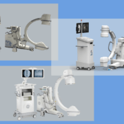
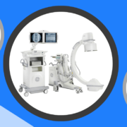
A C-Arm machine is a specialized medical imaging device that uses X-rays to produce real-time images of the body’s internal structures. Its name comes from the arm’s distinctive “C” shape, which allows the X-ray source and image intensifier to be positioned on opposite sides of the patient. This design provides unparalleled flexibility and maneuverability, enabling healthcare professionals to get high-quality images from virtually any angle.
What is an X-Ray Image Intensifier?
In any C-Arm fluoroscopy, the X-ray image intensifier is the central component that converts X-ray energy into visible light. The image intensifier produces top-quality images with minimal distortion and noise. A viewer can see real-time photos on a screen as the camera captures light. Some newer models now feature a flat detector instead of an image intensifier, as well.
Who Uses a C-Arm?
Many healthcare professionals use C-Arm machines, including orthopedic surgeons, interventional radiologists, and pain management specialists among many others. They are particularly well-suited for use during surgical procedures, as they allow doctors to visualize the area being treated with no invasive techniques. Some typical applications include:
- Orthopedic surgery, such as joint replacements or spinal fusions etc…
- Interventional radiology, including angioplasty and stent placement etc…
- Pain management for targeted injections, nerve blocks, kyphoplasty, RF Ablation, stimulator implants, etc…
What Are the Components of a C-Arm?
Several key components make up a C-Arm system, including:
- X-ray generator: This device generates X-rays to create images of the body’s internal structures.
- C-Arm: The characteristic “C” shaped arm houses the X-ray source and image intensifier , allowing them to be positioned opposite each other.
- Image intensifier: As discussed earlier, this component converts X-ray energy into visible light, which is then captured by a camera.
- Monitor: The monitor displays real-time images, enabling healthcare professionals to view and assess the patient’s condition during a procedure.
These are just a few of the essential components that make up a C-Arm system. With their incredible versatility and powerful imaging capabilities, these machines can be invaluable tools in the hands of an experienced healthcare provider.
If you want to upgrade your imaging equipment, contact us today, and let’s get started!
Reasons to Use a C-Arm System
There are several reasons a C-Arm fluoroscopy system is an invaluable tool for healthcare professionals:
- Real-time imaging: C-Arm machines provide real-time images, allowing doctors to make adjustments and confirm the success of a procedure as it’s happening.
- Minimally invasive: By providing clear, detailed images, C-Arm machines enable doctors to perform procedures with minimal invasiveness, reducing recovery time and improving patient outcomes.
- Versatility: A C-Arm machine’s unique design allows for unparalleled positioning flexibility, making it suitable for various applications.
- Efficiency: With high-quality images at the touch of a button, C-Arm machines streamline the imaging process, saving time and resources.
Ready to unlock the power of a C-Arm X-ray machine for your practice?
Contact ImagPros at in**@******os.com to learn more about our range of state-of-the-art imaging equipment and expert support services for C-Arm fluoroscopy. Together, we’ll ensure you have the tools to provide the best care for your patients.


Trackbacks & Pingbacks
[…] thorough guide uncovers the details about C-Arm fluoroscopy machines, addressing everything from their definition to their diverse components and […]
Comments are closed.